 |
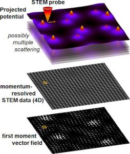
In this imaging mode diffraction patterns are recorded at each raster position of a scanning electron beam in an electron microscope. The result is a unique multidimensional data set that allows for the simultaneous evaluation and combination of real- and momentum space information. |
Applications of momentum-resolved STEM cover a broad spectrum: Combined with aberration-corrected electron optics, subatomic electric fields and charge densities can be measured down to a spatial resolution of 50pm in thin specimen. The access to the detailed angular distribution of scattered intensity opens new possibilities to optimise contrast dedicated to analyse distinct specimen properties independently, such as chemical composition, strain state, or specimen thickness. For soft matter, an biological specimen in particular, the ability to generate and optimise faint contrast in low-dose images in software after acquisition provides a high degree of flexibility and dose-efficiency. Momentum-resolved STEM has furthermore a large potential for the mapping of piezoelectric and spontaneous, as well as ferroelectric polarisations as a prerequisite to fundamentally understand and develop devices in the field of future information technology.
Within an international network spanning from Nebraska (USA) over the RWTH Aachen to Bangalore (India), the moreSTEM group at the Research Centre Jülich focuses on three main methodological and materials science challenges.